Methods and Materials
Preparation
Before beginning the experiment, sterile procedure must be followed. First wesocdyne was sprayed on a paper
towel and used to wipe down all surfaces involved in the experiment. Next, the
Oxford Bacti-cinerator was turned on and allowed to heat up. This took about 15
minutes.
Inoculation from a
broth
Per standard sterile procedure, inoculation should always be done
upside down to prevent microbial rain from infecting the specimen. When
inoculating from a bacterial broth it must first be vortexted. To do this, the
test tube was allowed to warm up to room temperature. Once at room temperature,
a loop was used to vortex the bacterial broth. Then it was wiped across the
nutrient agar plate and placed in an incubator at 30®C upside down to prevent
condensation from tampering with the growing bacteria.
Inoculation of
Bacteria
Several different bacterial samples were taken and will be used as the
independent variable in this experiment. Staphylococcus aureus beta-hemolytic,
staphylococcus aureus, mycobacteria, and the C. elegans food, Escherichia coli
(OP50) were inoculated on nutrient agar plates. These bacterias were inoculated
from test tubes containing bacterial broth. After inoculation they were placed
in an incubator for 48 hours at 30®C.
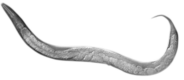
Caenorhabditis Elegans
Procedures
The C. elegans were picked from a stock plate using a spatula and a
Letica Zoom 2000 dissecting microscope. Using these tools, only stage four C.
elegans were picked off a stock plate and placed on a nutrient agar plate
containing their food source, OP50. Some plates were chunked using a standard
chunking apparatus. This was done buy pulling a small piece of agar off of the
agar plate and placing it on a fresh plate with a new supply of OP50. These
plates were then placed in an incubator indefinitely at 20®C.
Computer Software
A Dinolite camera was used to record the individual C. elegans once they
were on their own agar plate. After the C. elegans were picked, they had a 5-10
minute acclimation period to feed and explore their plate. After this gestation
period they were recorded at 10 minute interval through the Dinolite camera.
This camera was attached to a worm tracking software that can be used to
evaluate the behaviors of the C. elegans. However, the software was not
cooperating. Therefore, the video was saved and will be visually analyzed for
any strange behaviors.
Bleaching
Protocol
Using 5 M NaOH and household bleach (5% solution of sodium
hypochlorite) a plate of N2 C. elegans were bleached to extract the eggs. First
the C. elegan stock plates were washed in DI water 2 times to loosen the worms
and eggs stuck in the bacteria. The liquid was then sucked up in pipets
and put in a sterile test tube and capped. Then the bleaching solution was
pipeted in the test tubes, 0.5mL of 5 M NaOH and 1mL bleach. This was then
vortexed for approximately 30 seconds. After, the test tubes were centrifuged
for 30 seconds at 1000 g’s of force. The C. elegans were dead and the eggs left
at the bottom were put on a freshly seeded nutrient agar plate of OP50 using a
micropipette. The newly seeded plates were put in an incubator to grow at 20®C.
Chunking a
Plate
Using a dual-ended spatula/ chunking edge, the C. elegans were
chunked from a stock plate. This was done in the area closest to the center of
the concentration of OP50. After being chunked from the stock plate, the chunks
of C. elegans were placed in top side up on the new seeded plate of OP50. Then
placed into the incubator at 20®C.
Deseeding and Reseeding Agar
Plates
The OP50 plates were deseeded of their bacteria and reseeded with a new
bacteria used for the independent group studies. The plates were soaked in
wescodyne and allowed to sit for 15 seconds. Following that, they were scraped
with a loop to ensure the plate was completely clean. Then it was rinsed in
de-ionized water to wash off any remaining wescodyne.
Counting C.
elegans
Using a microscope and a colony counting
apparatus the worms on all plates were counted. The colony counting apparatus
was placed on the microscope and on top of that the agar plate. The
place was sectioned into quarters, and then counted a section at a time. One
quarter was counted, that number was multiplied by four to represent the total
number of worms on the plate.
